

The central nervous system consists of the brain, which is protected by the skull, and spinal cord, which is protected by the spine. The structure of the brain have various functions that are essential to life such as breathing, as well as our daily functioning, such as talking, walking, reading, and more. Structures of the brain are made up of the four parts which will be explained in detail below.
Basic Facts about the Human Brain
The structures of the brain are protected by the meninges (a membrane with three layers) and the skull. The soft, grayish-white brain weighs about three pounds and consists of two percent of one’s body weight. In fact, among the vertebrates, man has the largest brain size in proportion to his body size.
The brain has a rich blood supply from the arteries that deliver oxygen, glucose and other nutrients. However, it also has a blood-brain barrier that prevents certain substances like some drugs, from penetrating the brain.
Structures of the Brain: Cerebrum
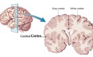
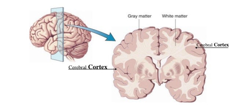
The Cerebral Cortex
The cerebrum consists of gyri (plural form of gyrus) or folded bulges that have deep furrows (fissures or sulci). It is a highly developed part where many mental functions originate, such as thinking and understanding language.
The cerebral cortex is functionally divided into lobes with specific tasks involved in thinking and reasoning, vision, hearing, movement, touch, and smell. Some functions, like language ability, are found in only one hemisphere.
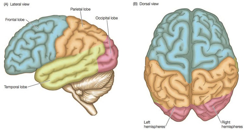
Four Lobes
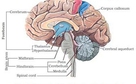
The right and left cerebral hemispheres are connected by a structure called corpus callosum. Each hemisphere has four lobes, each with different functions:
Parietal Lobe: involved in the reception and processing of sensory information from the body.
Frontal Lobe: responsible for planning, decision-making, and problem solving.
Temporal Lobe: responsible for memory, hearing, language, and emotion.
Occipital Lobe: responsible for vision.
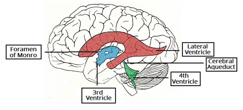
The Ventricles of the Brain
The structures of the brain that produce and transport the cerebrospinal fluid (CSF) are found in the ventricles. These are lined by special ependymal cells, which form the choroid plexus where CSF is produced.
There are four ventricles in the brain: right lateral and left lateral, and the third and fourth ventricles.
Lateral Ventricles
The lateral ventricles are located in the left and right hemispheres, with ‘horns’ projecting into the frontal, temporal, and occipital lobes. The lateral ventricles increase in volume as you age.
Third Ventricle
The right and left lateral ventricles are connected to the 3rd ventricle through the foramen of Monro. This ventricle is found between the left and right thalamus. It has two protrusions, consisting of the supra-optic recess, which is above your optic chiasm, and the infundibular recess, which is above your optic stalk.
Fourth Ventricle
This structure, which is found in the brainstem, receives CSF from your third ventricle through the cerebral aqueduct. From here, CSF drains into the central spinal canal, where it bathes the spinal cord, and into the subarachnoid cisterns, where it bathes the brain. CSF is reabsorbed from the arachnoid and pia mater and goes back into circulation.
Structures of the Brain: Diencephalon
The diencephalon and the cerebrum (telencephalon) make up the forebrain (prosencephalon). Main structures of the brain that make up the diencephalon comprise the thalamus, epithalamus (and pineal gland), hypothalamus, and subthalamus. It relays the sensory information and controls autonomic functions of your peripheral nervous system.
The diencephalon also joins the endocrine and the nervous system structures and coordinates with the limbic system to manage memories and emotions.
The thalamus has connections with the cerebral cortex and spinal cord. It processes sensory perception and regulates of motor functions.
The hypothalamus is a small but vital component of the brain. It takes part in the regulation of hormones, body temperature, and many other activities.
The epithalamus is in the most dorsal portion of the diencephalon. It contains an organ called the pineal gland, which secretes melatonin.
The limbic system consists of primitive brain structures found at the top of the brainstem under the cortex. These structures are involved in motivations and emotions like fear, anger, pleasure and those relating to sexual behavior. Two large structures called the hippocampus and amygdala play significant roles in memory function.
Structures of the Brain: Brain Stem
The brainstem connects the cerebrum to the spinal cord. It is made up of the midbrain, the medulla oblongata, and pons. Motor and sensory signals are relayed between the brain and the spinal cord through the brainstem. It also controls autonomic functions which support life.
The mesencephalon (midbrain) connects the hindbrain to the forebrain.
The medulla oblongata controls autonomic functions like digestion, breathing, swallowing, sneezing, and blood vessel and heart function. It also relays motor and sensory signals from the midbrain to the forebrain. Being part of the brainstem, it helps transfer messages between the brain and spinal cord.
The pons (“bridge”) is part of the hindbrain that connects the medulla oblongata with the cerebral cortex. It serves as a coordination center between the cerebral hemispheres. It helps transfer messages between the brain and spinal cord.
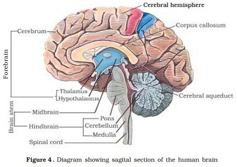
Structures of the Brain: Cerebellum
Cerebellum, in Latin, means “little brain”. It is the part of the hindbrain that coordinates motor movement, balance, and muscle tone. Just like the cerebrum, it is composed of white and gray matter. Its outer layer has smaller, compact folds compared to those in the cerebral cortex. It has millions of neurons involved in processing data and relaying information between the cerebral cortex and the muscles involved in motor control.
Watch this video to learn more about the basic parts of the brain.
View All Comments /Add Comment